


聯(lián)系電話(huà)
0797-5591768
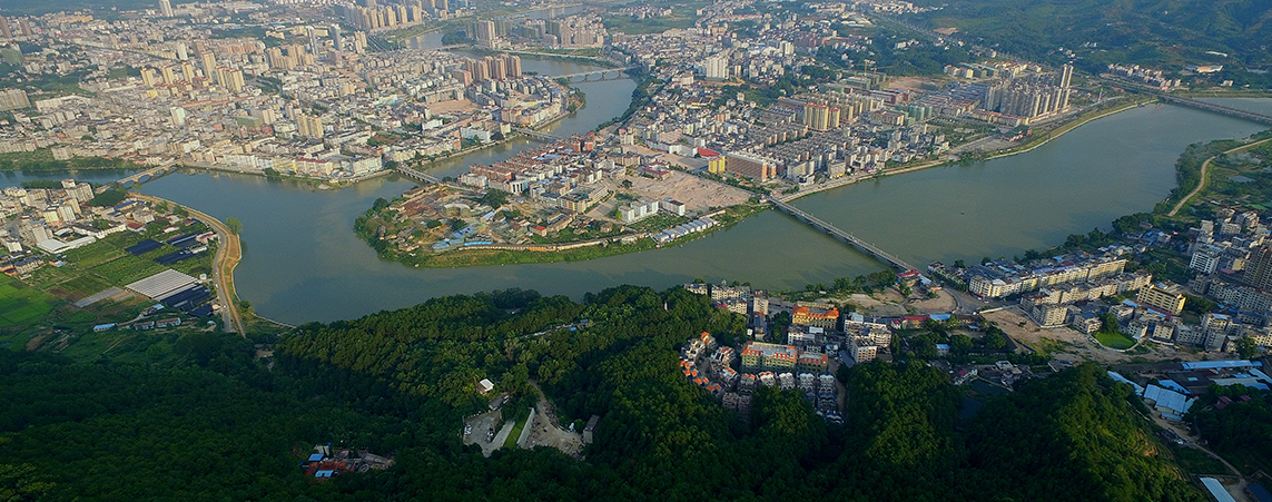
 會(huì)昌縣冷鏈物流倉(cāng)儲(chǔ)中心招商公告
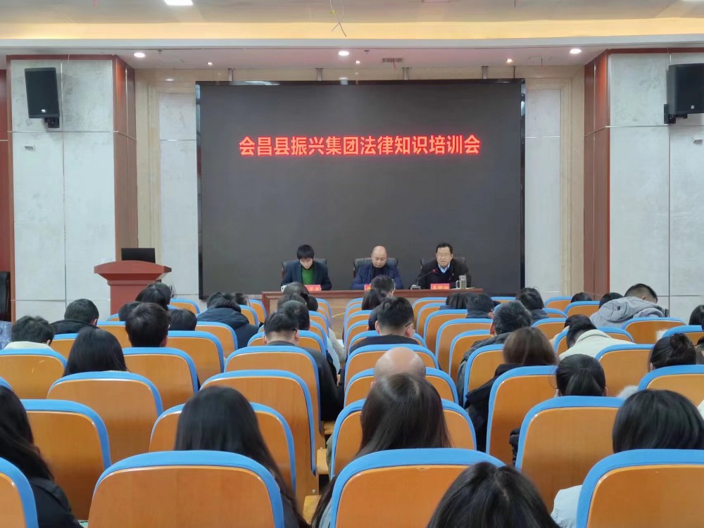
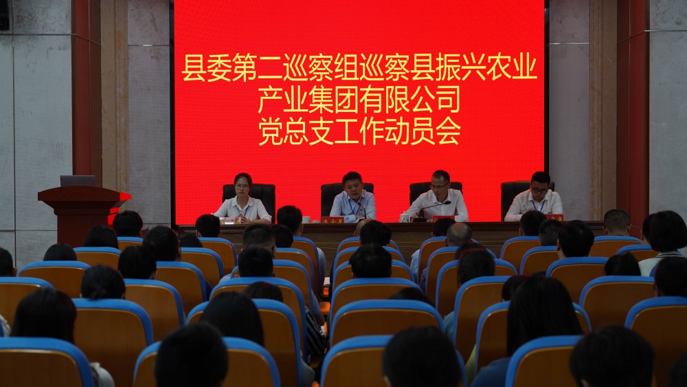
中共會(huì)昌縣振興農(nóng)業(yè)產(chǎn)業(yè)集團(tuán)有限...
會(huì)昌縣贛南肉牛產(chǎn)業(yè)綜合交易市場(chǎng)...
會(huì)昌縣至簡(jiǎn)數(shù)字產(chǎn)業(yè)有限公司招聘...
關(guān)于會(huì)昌縣工業(yè)園區(qū)標(biāo)準(zhǔn)廠(chǎng)房(N...
會(huì)昌縣融合項(xiàng)目管理有限公司建設(shè)...
會(huì)昌縣融合項(xiàng)目管理有限公司建設(shè)...
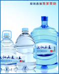
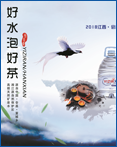
江西園山食品飲料有限責(zé)任公司 ...
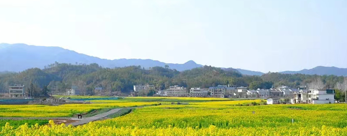
關(guān)于公開(kāi)征集會(huì)昌縣白鵝鄉(xiāng)示范鎮(zhèn)...
會(huì)昌縣冷鏈物流倉(cāng)儲(chǔ)中心招商公告
中共會(huì)昌縣振興農(nóng)業(yè)產(chǎn)業(yè)集團(tuán)有限...
會(huì)昌縣贛南肉牛產(chǎn)業(yè)綜合交易市場(chǎng)...
會(huì)昌縣至簡(jiǎn)數(shù)字產(chǎn)業(yè)有限公司招聘...
關(guān)于會(huì)昌縣工業(yè)園區(qū)標(biāo)準(zhǔn)廠(chǎng)房(N...
會(huì)昌縣融合項(xiàng)目管理有限公司建設(shè)...
會(huì)昌縣融合項(xiàng)目管理有限公司建設(shè)...
江西園山食品飲料有限責(zé)任公司 ...
關(guān)于公開(kāi)征集會(huì)昌縣白鵝鄉(xiāng)示范鎮(zhèn)...